

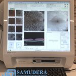

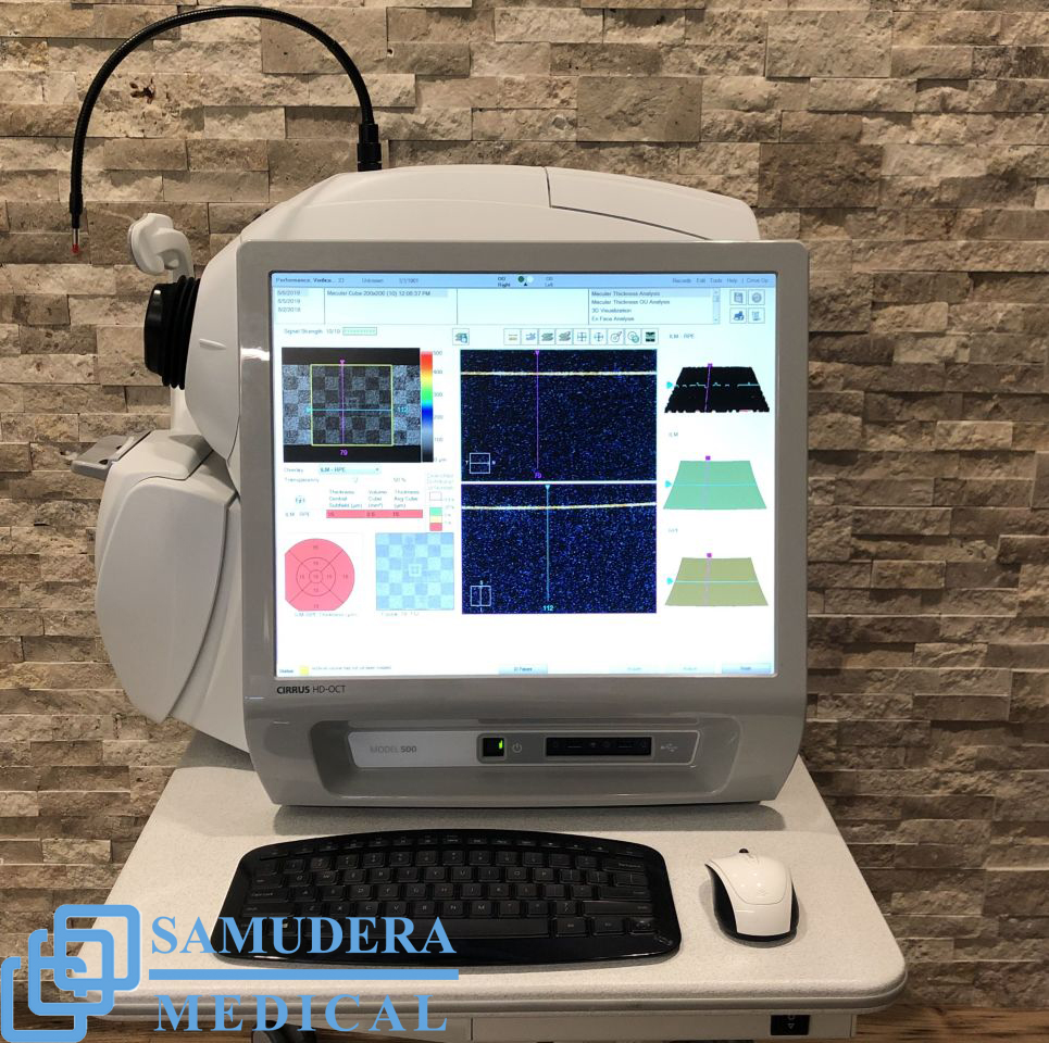
Sale – Carl Zeiss Cirrus 500 HD OCT
Original price was: $9,800.$6,850Current price is: $6,850.
CompareDescription
Carl Zeiss Cirrus 500 HD OCT
Carl Zeiss Cirrus 500 HD OCT Electrical and Physical
Weight: 76 lbs (34 kg)
Dimensions of instrument: 26L x 18W x 21H (in), 65L x 46W x 53H (cm)
Dimensions of table: 39L x 22W (in), 99L x 56W (cm)
Fixation: Internal, external
Internal fixation focus adjustment: -20D to +20D (diopters)
Electrical rating (115V): Single Phase, 100–120V~ systems: 50/60Hz, 5A
Electrical rating (230V): Single Phase, 220–240V~ systems: 50/60Hz, 2.5A
Carl Zeiss Cirrus 500 HD OCT
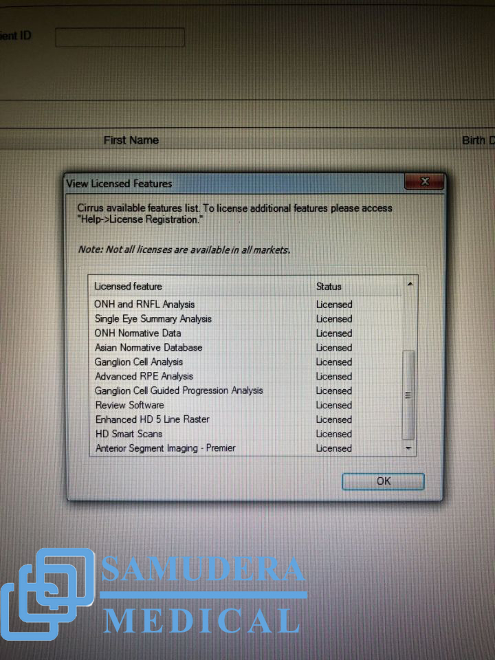
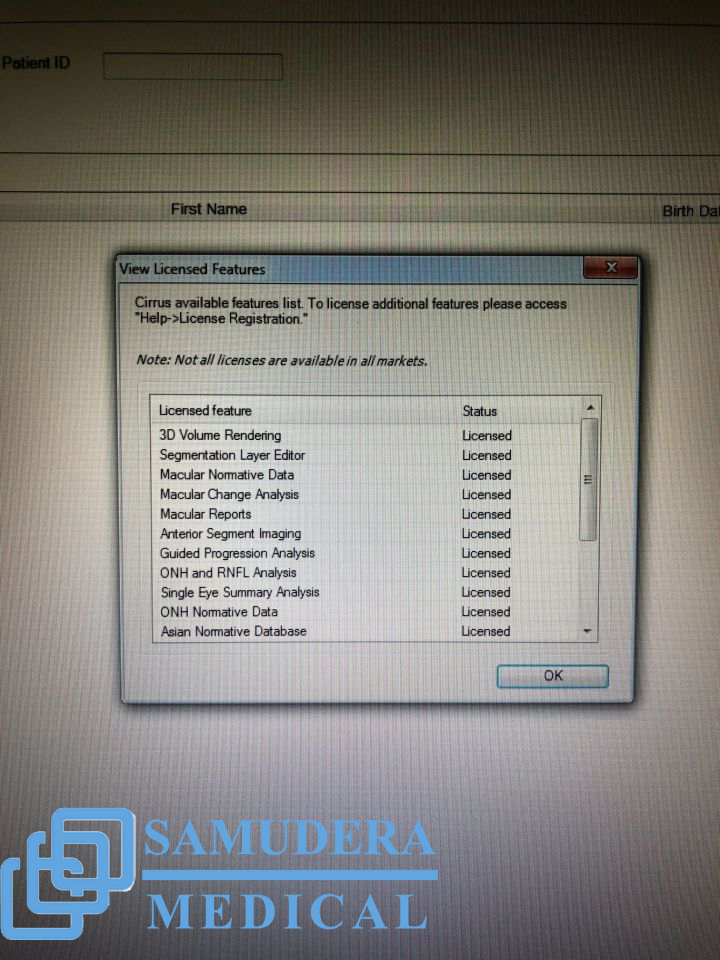
* Macular Thickness OU Analysis
* Ganglion Cell Analysis
* Guided Progression Analysis (GPA™)
* Macular Thickness and Change Analysis
* Macular Thickness Normative Data
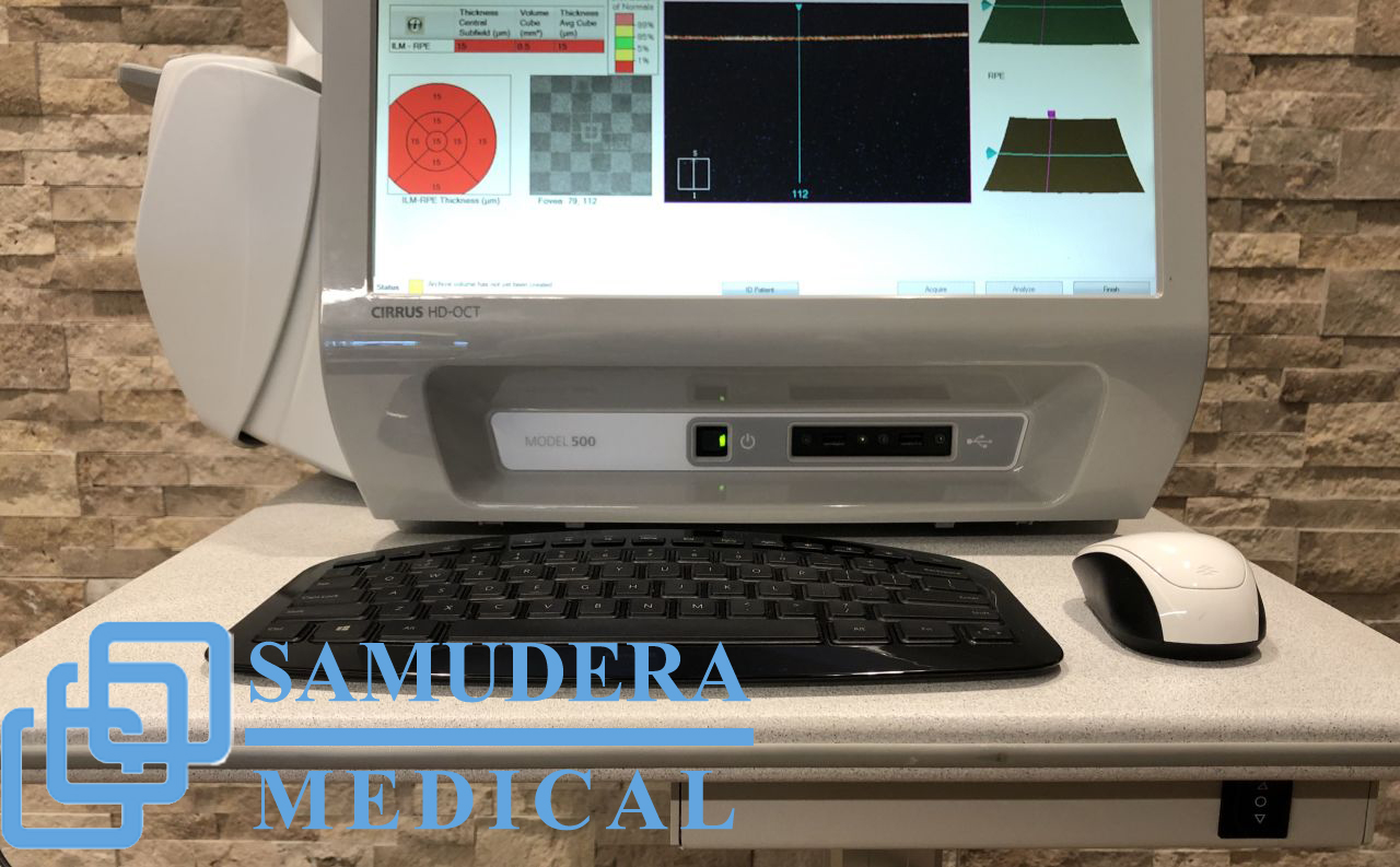
The Essential OCT CIRRUS HD-OCT 500
Offers comprehensive care practices essential OCT capabilities with a broad range of clinical applications in an easy-to-learn, easy-to-use instrument. It aids in the management of glaucoma and retinal disease, retina assessment for cataract surgery and anterior segment imaging for corneal disease.
Examine Retinal Details: Selective Pixel Profiling optimizes each illumination point in the 20 X HD Raster Scan, ensuring detail-rich visualizations that spotlight critical pathological elements.
Gain Deeper Insight: New Enhanced Depth Imaging focuses the signal lower in the scan window for assessment of the deeper choroidal tissue.
Visualize Change: New FastTrac on the CIRRUS HD-OCT 5000 precisely targets and captures the same tissue every time to ensure consistent comparison.
Carl Zeiss Cirrus 500 HD OCT Insightful Analyses
Reproducible Analyses: Zeiss propietary algorithms measure and display layers for unsurpassed tissue targeting, segmentation, and reproducible measurements.
Comparative References: Diversified normative databases of ONH, RNFL, Ganlion Cell / IRL, and Macular Thickness facilitate at-a-glance identification of anatomy outside normal limits.
Change Measurements: All Cirrus data cubes are automatically registered with historical data, allowing for point-to-point measurements of change.
OCT Imaging
Methodology: Spectral domain OCT
Optical source: Superluminescent diode (SLD), 840 nm
Scan speed: 27K- 68K A-scans per second
A-scan: 2.0 mm (in tissue), 1024
Axial resolution: 5 μm (in tissue)
Transverse resolution: 15 μm (in tissue)
Carl Zeiss Cirrus 500 HD OCT Fundus Imaging
Methodology: Love OCT Fundus
Live fundus image: During alignment
Optical source: Superluminescent diode (SLD), 840 nm
Field of view: 36 degrees W x 30 degrees H
Frame rate: > 1.7 Hz
Transverse resolution: 45 μm (in tissue)
Carl Zeiss Cirrus 500 HD OCT Iris Imaging
Methodology: CCD camera
Resolution: 1280 x 1024
Live iris image: During alignment
Related products
-
- Sale!
- Ophthalmology Equipment, Zeiss
Sale – Carl Zeiss Cirrus 4000 HD OCT
- Original price was: $13,000.$7,950Current price is: $7,950.
- Add to cart
-
- Sale!
- Essilor, Ophthalmology Equipment
Sale – Essilor Gamma Patternless Edger
- Original price was: $6,900.$4,250Current price is: $4,250.
- Add to cart
-
- Sale!
- Autorefractor, Keratometer, Nidek, Ophthalmology Equipment
Sale – Nidek AFC-330 Auto Fundus Retinal Camera Complete
- Original price was: $7,500.$4,950Current price is: $4,950.
- Add to cart


Reviews
There are no reviews yet.