


Sale – Carl Zeiss Cirrus 4000 HD OCT
Original price was: $13,000.$7,950Current price is: $7,950.
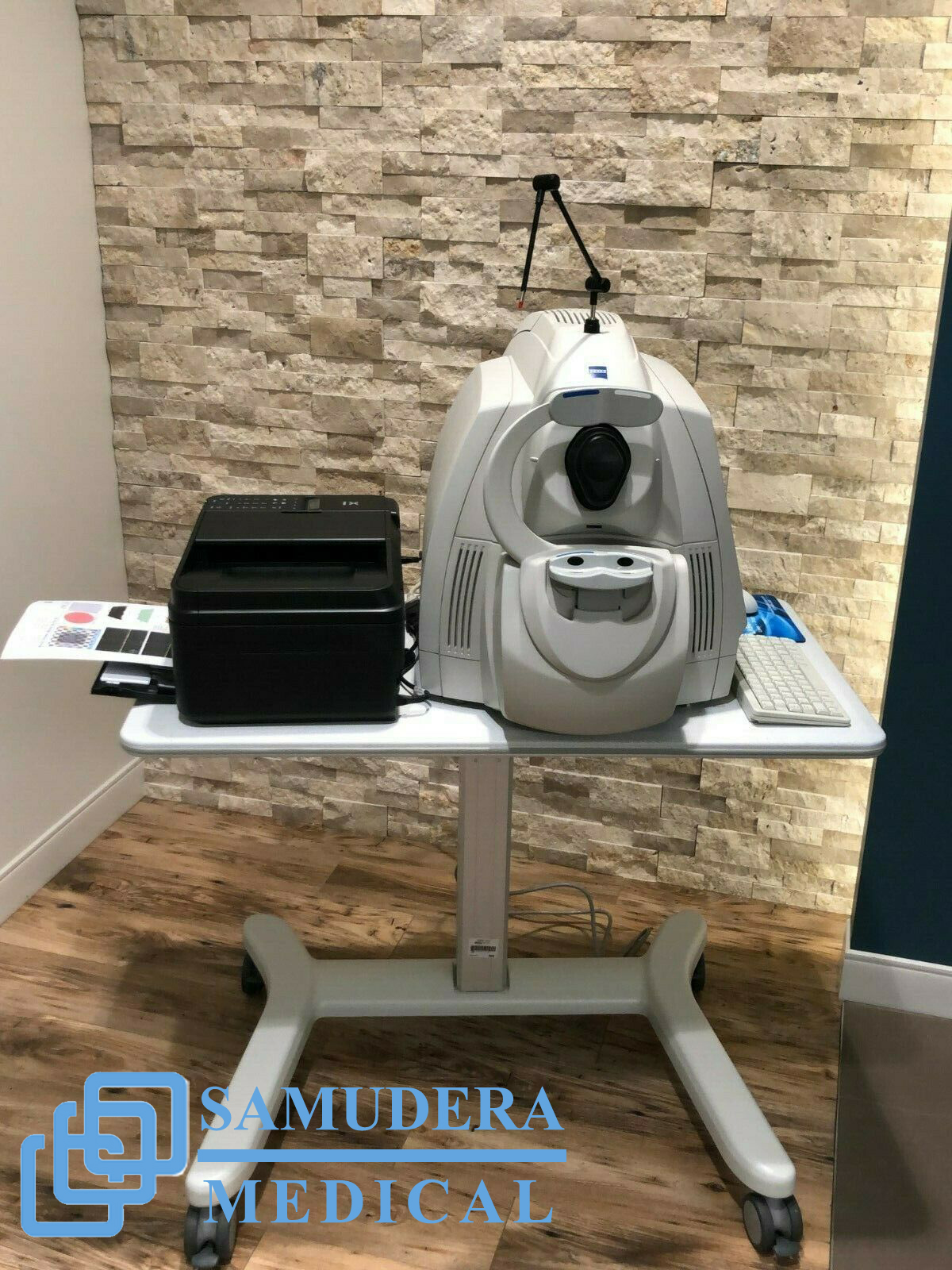
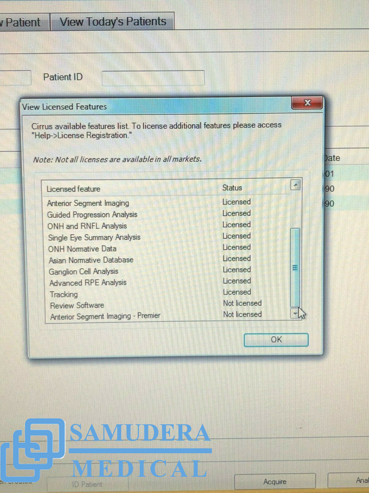
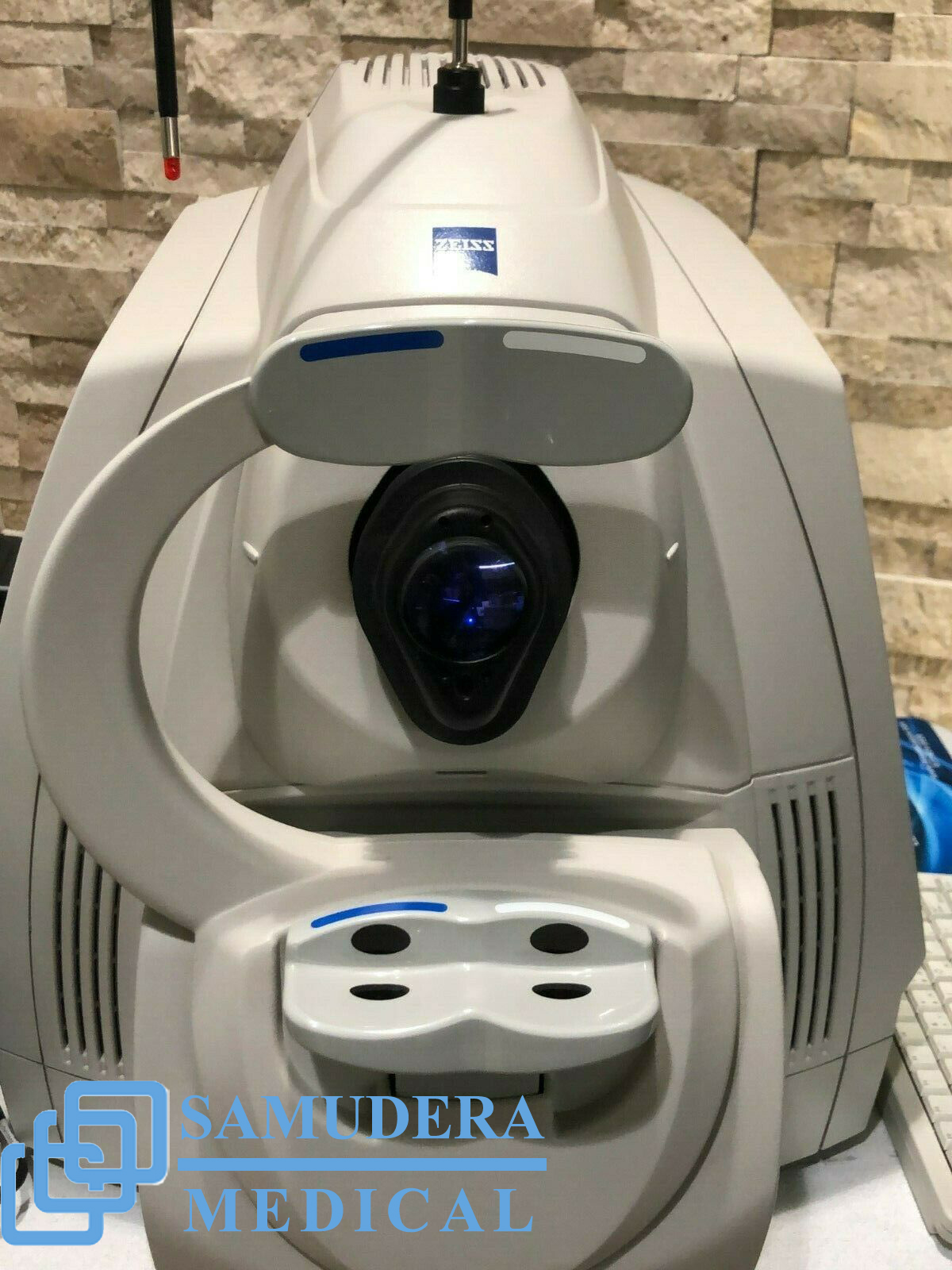
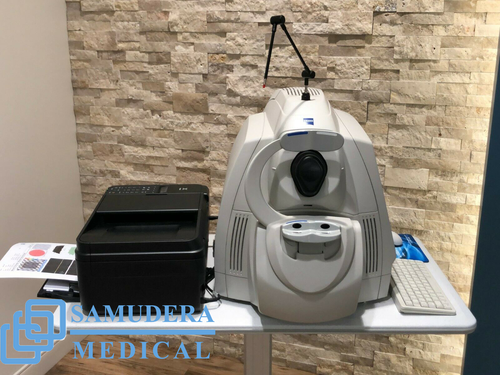
CARL ZEISS Cirrus HD-OCT 4000 analyses glaucoma and retina with modern integrated design, ease of use, and small footprint.
Description
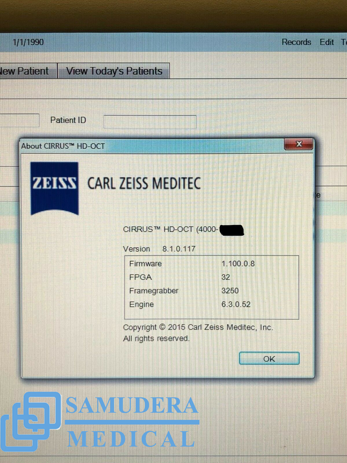
Carl Zeiss Cirrus 4000 HD OCT
Carl Zeiss Cirrus 4000 HD OCT KEY FEATURES
Built on 10 years experience at the vanguard of innovation, Carl Zeiss Meditec OCT technology has become the recognized standard of care. Now, Cirrus HD-OCT offers another leap forward with a superior platform that delivers unprecedented imaging details for clinical decision making.
ZEISS optics provide superior visualization of anatomical details across a wider range of patients.
Robust engineering with premium components ensures consistent precision performance.
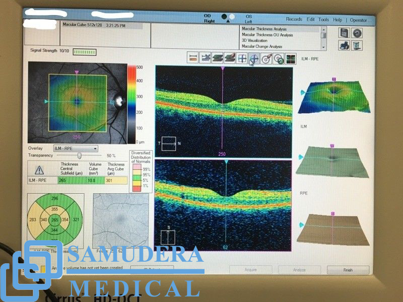
Unique HD layer maps and images highlight clinically relevant details for identification and monitoring of specific diseases – all at a glance.
The powerful Cirrus HD-OCT scan engine delivers superior image data. The new HD Enhanced Raster Scan leverages this power to produce images with outstanding detail while maintaining patient throughput. The proprietary Selective Pixel Profiling technology enhances anatomical features while reducing image noise.
Cirrus HD-OCT enables repeatable visualization of clinically relevant anatomy with exact correlation between the OCT scan and the fundus image. Comprehensive navigational tools ensure efficient and simple operation.
Carl Zeiss Cirrus 4000 HD OCT Designed for efficiency
Small footprint and integrated design are ideal for crowded or busy practice
90 degree orientation facilitates observation of patient throughout exam
Advanced optics aid in the examination of patients with cataracts
Dilation is not required even for pupils as small as 2.5 mm
Mouse Driven Alignment™ delivers superior image capture and analysis in just a few clicks, resulting in reduced chair time for the patient
Auto Patient Recall™ assures patient position and instrument setting are repeated from previous visit
Carl Zeiss Cirrus 4000 HD OCT Technical data
OCT Scanning
Axial resolution: 5 μm (in tissue)
Transverse resolution: 15 μm (in tissue)
Scan speed: 27,000 A-scans per second
A-scan depth: 2.0 mm (in tissue), 1024 points
Optical source: superluminescent diode (SLD), 840 nm
Fundus Imaging
Line scanning ophthalmoscope (LSO)
Live during scanning
Transverse resolution: 25 μ (in tissue)
Optical source: superluminescent diode (SLD), 750 nm
Field of view: 36° x 30°
Scan Patterns
Macular Cube 200 x 200 Combo: 200 horizontal scan lines comprised of 200 A-scans
Macular Cube 512 x 128 Combo: 128 horizontal scan lines comprised of 512 A-scans
5 Line Raster: 4096 A-scans per B-Scan (adjustable length, spacing and orientation)
Focus Adjustment Range
−20D to +20D (diopters)
Fixation
Internal and external
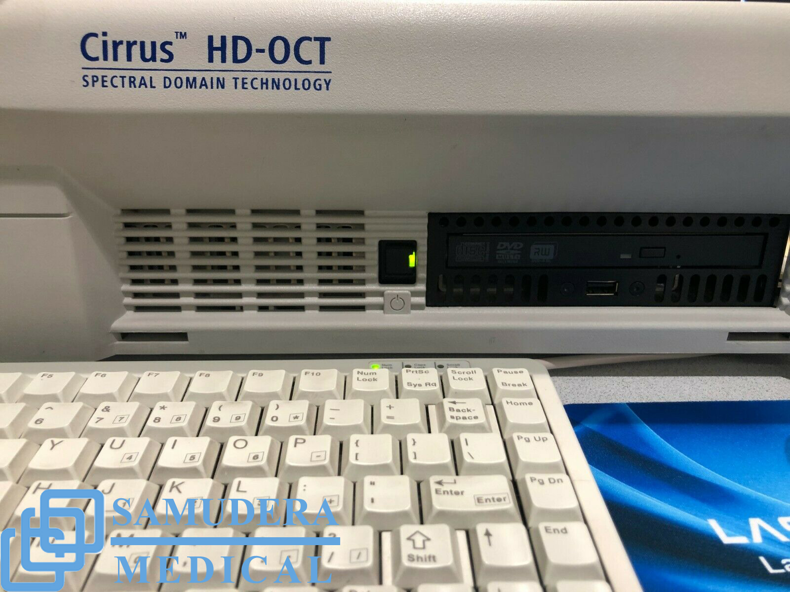
Computer
Windows 7 OS
Internal storage: > 80,000 scans
CD-RW, DVD-ROM drive
Integrated 15″ color flat panel display
Pupil Size Requirement
≤ 2.0 mm (≥ 3.0 mm optimal for LSO)
Dimensions/Weight (Instrument Only)
25.6 L x 17.3 W x 20.9 H (in); 65 L x 44 W x 53 H (cm)
83 lbs; 37.6 kg
Electrical
100-120V~, 50/60Hz, 5A 220-240V~, 50/60Hz, 2.5A
CARL ZEISS Cirrus HD-OCT 4000 analyses glaucoma and retina with modern integrated design, ease of use, and small footprint.
With the smaller budget designed Cirrus HD-OCT 4000 focused on the essential core OCT functionality.CARL ZEISS Cirrus HD-OCT Have two models, Model 400 and Model 4000 which capable of anterior segment imaging and offer the same package of glaucoma and retina analyses
Carl Zeiss Cirrus 4000 HD OCT allows visualize and analyze patient’s condition and captures a tightly packed, detail-rich cube of data in just seconds and make this unit as best-selling spectral domain OCT system
Computer
• High-performance multi-core processor
• Internal storage: > 80,000 scans
• CD-RW, DVD-ROM drive
• Integrated 15″ color flat panel display
INCLUDES
-Scanning system Live OCT Fundus Technology
-Field of view 36 x 22 degrees
-Methodology Spectral domain
-Scan speed 27,000 A-scans / sec
-A-scan depth 2.0 mm (in tissue)
-Axial resolution 5 µm (in tissue)
-Transverse resolution 15 µm (in tissue)
-Processor Intel Pentium Core 2 Duo, Win XP
Related products
-
- Sale!
- Autorefractor, Keratometer, Nidek, Ophthalmology Equipment
Sale – Nidek AFC-330 Auto Fundus Retinal Camera Complete
- Original price was: $7,500.$4,950Current price is: $4,950.
- Add to cart
-
- Sale!
- Essilor, Ophthalmology Equipment
Sale – Essilor Neksia Lens Edging System
- Original price was: $8,500.$6,250Current price is: $6,250.
- Add to cart
-
- Sale!
- Ophthalmology Equipment
Sale – Optos California fa Ultra-widefield Retinal Imaging Systems
- Original price was: $17,800.$12,750Current price is: $12,750.
- Add to cart


Reviews
There are no reviews yet.